Conjoint Tendon Shoulder Anatomy / Conjoined Tendon Shoulder Anatomy - Capsular Attachment Of ... : It reduces wear and tear on the tendon during movement at the shoulder.
Conjoint Tendon Shoulder Anatomy / Conjoined Tendon Shoulder Anatomy - Capsular Attachment Of ... : It reduces wear and tear on the tendon during movement at the shoulder.. The conjoint tendon then turns inferiorly and attaches on. Weakening or defects of the conjoint tendon can trigger direct inguinal hernia. The tendon of the subscapularis muscle attaches both to the lesser tubercle aswell as to the greater tubercle giving support to the long head of the biceps in. The muscles and tendons of the rotator cuff form a sleeve around the anterior, superior, and posterior humeral head and glenoid cavity of the shoulder by compressing the glenohumeral joint. Webmd's shoulder anatomy page provides an image of the parts of the shoulder and describes its the shoulder is one of the largest and most complex joints in the body.
Robin smithuis and henk jan van der woude. Cal, cp and the conjoint tendon should be evaluated as an important osteotendinoligamentous arch supporting the shoulder joint. In this episode of eorthopodtv, orthopaedic surgeon randale c. Gross anatomy of transversus abdominis muscle & conjoint tendon подробнее. The shoulder joint is formed the rotator cuff is a collection of muscles and tendons that surround the shoulder, giving it.
Dislocated Shoulder | Cleveland Clinic from www.clevelandclinic.org Kaddress correspondence to robert f. Shoulder radiology & anatomy at usuhs.mil. Joint via its conjoint tendon, the achilles tendon. The conjoint tendon was released from fascial attachments to the capsule to mobilize the. Shoulder muscles and shoulder tendons. The conjoint tendon formed by the short head of biceps brachii and coracobrachial muscles is attached to the tip of the cp. The conjoint tendon, also known as henle's ligament, forms when the medial fibers of the internal oblique aponeurosis unite with the deeper fibers of the transversus abdominis aponeurosis. Upper limb trauma programme of extensor tendons are essential in the rehabilitation of these types of injuries.
Tendon transfers around the shoulder подробнее.
Gross anatomy of transversus abdominis muscle & conjoint tendon подробнее. Kaddress correspondence to robert f. Changes in neurovascular anatomy after open latarjet procedure 3. Muscles allow us to move by pulling on bones. Qualitative and quantitative anatomy of the proximal. There are several important ligaments in the shoulder. Joint via its conjoint tendon, the achilles tendon. Ligaments are soft tissue structures that connect bones to bones. Coracoid process, component of conjoint tendon insertion: Related online courses on physioplus. Shoulder muscles and shoulder tendons. The conjoint tendon was released from fascial attachments to the capsule to mobilize the. • during abduction of the shoulder joint, the supraspinatus tendon is exposed to friction against the acromion.
Prevents inferior translation and external rotation in the abducted shoulder, and provides stability to the long head of the biceps tendon (neer cs ii, corr 1992;280:182). The abdominal wall is split into the posterior (back), lateral (sides). The long head of biceps (lhb) is a very important tendon that travels through the shoulder joint (glenohumeral joint). • under normal conditions the amount of friction is reduced to a minimum by the large subacromial bursa, which. These are the main ligaments that help to stabilize the joints of.
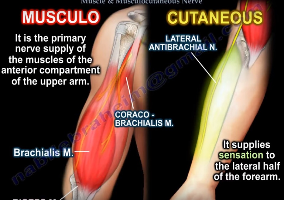
Anatomy of the Coracobrachialis and the Musculocutaneous ... from orthopaedicprinciples.com Tendon conjoint — le tendon conjoint ici noté inguinal aponeurotic falx le tendon conjoint est une structure fibreuse constitué de la réunion des terminaisons fibreuses des muscles oblique interne et transverse de l abdomen. It reduces wear and tear on the tendon during movement at the shoulder. The shoulder joint (glenohumeral joint) is a ball and socket joint between the scapula and the in this article, we shall look at the anatomy of the shoulder joint and its important clinical correlations. Muscles allow us to move by pulling on bones. Для просмотра онлайн кликните на видео ⤵. The long head of biceps (lhb) is a very important tendon that travels through the shoulder joint (glenohumeral joint). An image depicting shoulder anatomy can be seen below. Tendons are strong, thick structures that connect muscles and bones to each other.
Related online courses on physioplus.
Normal mri anatomy of the musculoskeletal system. Kaddress correspondence to robert f. Schematic representation of the right shoulder. Sechrest, md narrates an animated tutorial on the basic anatomy of the shoulder. Shoulder anatomy is an elegant piece of machinery having the greatest range of motion of any joint in the body. • during abduction of the shoulder joint, the supraspinatus tendon is exposed to friction against the acromion. Related online courses on physioplus. The shoulder | musculoskeletal key. These are the main ligaments that help to stabilize the joints of. Normal anatomy, variants and checklist. Shoulder radiology & anatomy at usuhs.mil. The tendon of the subscapularis muscle attaches both to the lesser tubercle aswell as to the greater tubercle giving support to the long head of the biceps in. The conjoint tendon was released from fascial attachments to the capsule to mobilize the.
The conjoint tendon (previously known as the inguinal aponeurotic falx) is a sheath of connective tissue formed from the lower part of the common aponeurosis of the abdominal internal oblique muscle and the transversus abdominis muscle, joining the muscle to the pelvis. Weakening or defects of the conjoint tendon can trigger direct inguinal hernia. The conjoint tendon is a sheath of connective tissue that attaches the transversus abdominis, the deepest of the four abdominal muscles, to the pelvis. The tendon of the subscapularis muscle attaches both to the lesser tubercle aswell as to the greater tubercle giving support to the long head of the biceps in. Schematic representation of the right shoulder.
Module 1: Lecture 13 - Inguinal Region of Anterior ... from o.quizlet.com These are the main ligaments that help to stabilize the joints of. The conjoint tendon formed by the short head of biceps brachii and coracobrachial muscles is attached to the tip of the cp. Changes in neurovascular anatomy after open latarjet procedure 3. Kaddress correspondence to robert f. Normal anatomy, variants and checklist. In this episode of eorthopodtv, orthopaedic surgeon randale c. Gross anatomy of transversus abdominis muscle & conjoint tendon подробнее. The shoulder | musculoskeletal key.
In this episode of eorthopodtv, orthopaedic surgeon randale c.
An image depicting shoulder anatomy can be seen below. Для просмотра онлайн кликните на видео ⤵. The conjoint tendon was released from fascial attachments to the capsule to mobilize the. The biceps muscle has two tendons at the shoulder, called the long head and short head. Sechrest, md narrates an animated tutorial on the basic anatomy of the shoulder. Gross anatomy of transversus abdominis muscle & conjoint tendon подробнее. Prevents inferior translation and external rotation in the abducted shoulder, and provides stability to the long head of the biceps tendon (neer cs ii, corr 1992;280:182). Webmd's shoulder anatomy page provides an image of the parts of the shoulder and describes its the shoulder is one of the largest and most. In this episode of eorthopodtv, orthopaedic surgeon randale c. • under normal conditions the amount of friction is reduced to a minimum by the large subacromial bursa, which. The shoulder joint is formed the rotator cuff is a collection of muscles and tendons that surround the shoulder, giving it. Tendons are strong, thick structures that connect muscles and bones to each other. Changes in neurovascular anatomy after open latarjet procedure 3.
Qualitative and quantitative anatomy of the proximal shoulder tendon anatomy. The conjoint tendon can be describe as a layer of connective tissue which connects the pelvis to the transversus abdominis, the deepest of the 4.


0 Komentar